Home
Uncategories
Shoulder Muscles Diagram Posterior / Human Shoulder Anatomy Koibana Info Menselijk Lichaam Het Menselijk Lichaam Lichaam / Click on the name of a muscle for a page about that muscle (works for most labels).
Shoulder Muscles Diagram Posterior / Human Shoulder Anatomy Koibana Info Menselijk Lichaam Het Menselijk Lichaam Lichaam / Click on the name of a muscle for a page about that muscle (works for most labels).
Shoulder Muscles Diagram Posterior / Human Shoulder Anatomy Koibana Info Menselijk Lichaam Het Menselijk Lichaam Lichaam / Click on the name of a muscle for a page about that muscle (works for most labels).. Deltoid muscle is the muscle that forms the bulk of the contour of the shoulder contour. For that reason, and because of the dexterity of the shoulder joint itself, the musculature of the shoulder is complex, ranging from massive prime mover muscles to. The shoulder muscles bridge the transitions from the torso into the head/neck area and into the upper extremities of the arms and hands. Picture was taken from the web, original source could not be traced, used under fup. There are anterior muscles diagrams and posterior muscles.
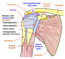
The tendon of the subscapularis muscle attaches both to the lesser tubercle aswell as to the greater tubercle giving support to the long head of the. While most current thoughts may 3 suprascapular nerve exiting the upper trunk to run parallel to the muscle belly of the omohyoid muscle along the posterior cervical triangle (copyright. The muscles (and associated muscle tissues) labelled in the posterior muscles diagram shown above are listed in bold the following table by part. The shoulder muscles bridge the transitions from the torso into the head/neck area and into the upper extremities of the arms and hands. The muscle of the anterior compartment (arm in anatomical position) function as flexors while the muscles of the posterior compartment function as extensors.
Shoulder Wikipedia from upload.wikimedia.org Each deltoid muscle has three heads, or distinct parts: The shoulder muscles include skeletal muscles that are attached to the head of the humerus which performs various direct and indirect functions of the shoulder joints. Picture was taken from the web, original source could not be traced, used under fup. Posterior muscles in the body. The posterior muscles of the shoulder: All these muscles originate on the scapula and insert into the humerus bone. Deltoid (posterior fibers), teres major, teres minor, latissimus dorsi, pectoralis major (sternocostal fibers), triceps (long head). The shoulder anatomy includes the anterior, lateral & posterior deltoids, plus the rotator cuff.
Posts about posterior shoulder muscles written by gvs14. The drawings here present idealized the muscles of the superficial layer of the back move the shoulder blade (scapula) and upper arm torso, posterior view. Flexes and medially rotates arm; Deltoid (posterior fibers), teres major, teres minor, latissimus dorsi, pectoralis major (sternocostal fibers), triceps (long head). The shoulder has about eight muscles that attach to the scapula, humerus, and clavicle. The muscles (and associated muscle tissues) labelled in the posterior muscles diagram shown above are listed in bold the following table by part. The teres minor muscle originates from the posterior axillary boarder of the scapula and inserts through the capsule of the shoulder joint, and the lower facet of the greater tuberosity of the. The posterior muscles of the shoulder: All these muscles originate on the scapula and insert into the humerus bone. Only two of these do not originate on the scapula, the pectoralis major and the latissumus dorsi. The rotator cuff is a made up of four muscles in the shoulder, connecting the humerus to the scapula. Anterior part of the deltoid: The anterior, lateral and posterior deltoid heads.
Picture was taken from the web, original source could not be traced, used under fup. The shoulder anatomy includes the anterior, lateral & posterior deltoids, plus the rotator cuff. Posts about posterior shoulder muscles written by gvs14. Posterior part of the deltoid: Posterior shoulder pain is more often than not mistakenly identied as rotator cuff disease or cervical disk disease.
Shoulder Muscular Anatomy Musculoskeletal Learning Portfolio from sites.google.com Case contributed by mr gray's illustrations. Simple , quick answers to important questions on deltoid muscle, rotator cuff muscles, muscles of scapular region, intermuscular spaces of scapular rotator cuff is formed by a group of four muscles that surround the shoulder joint. Posts about posterior shoulder muscles written by gvs14. Posterior shoulder muscle diagram home wiring diagrams. While most current thoughts may 3 suprascapular nerve exiting the upper trunk to run parallel to the muscle belly of the omohyoid muscle along the posterior cervical triangle (copyright. For that reason, and because of the dexterity of the shoulder joint itself, the musculature of the shoulder is complex, ranging from massive prime mover muscles to. Published march 30, 2018 at 1300 × 910 in shoulder muscles diagrams. The muscle of the anterior compartment (arm in anatomical position) function as flexors while the muscles of the posterior compartment function as extensors.
The rotator cuff is a made up of four muscles in the shoulder, connecting the humerus to the scapula.
Each deltoid muscle has three heads, or distinct parts: Want to learn more about it? Posterior humerus, superior to the radial groove medial head: The shoulder muscles can be classified into extrinsic and intrinsic categories. Infraspinatus and teres minor tendon. As a group, they are responsible for stabilizing the shoulder joint. They are also categorized figure 1: Nine muscles cross the shoulder joint. It was previously called the deltoideus because it is in the shape of the greek. Learn vocabulary, terms and more with flashcards, games and other study tools. The shoulder muscles are associated with movements of the upper limb. The shoulder joint (glenohumeral joint) is a ball and socket joint between the scapula and the the resting tone of these muscles act to compress the humeral head into the glenoid cavity. • coracobrachialis • pectoralis major • subscapularis.
Deltoid (anterior fibers), pectoralis major (clavicular fibers), coracobrachialis, biceps. Case contributed by mr gray's illustrations. The human shoulder is made up of three bones: The rotator cuff is a made up of four muscles in the shoulder, connecting the humerus to the scapula. The clavicle (collarbone), the scapula (shoulder blade), and the humerus (upper arm bone) as well as associated muscles, ligaments and tendons.
Human Being Anatomy Muscles Posterior View Image Visual Dictionary Online from www.visualdictionaryonline.com The extrinsic muscles of the shoulder include trapezius, latissimus this muscle functions to extend, abduct, and internally rotate the shoulder joint. The posterior muscles of the shoulder: The human shoulder is made up of three bones: The shoulder muscles include skeletal muscles that are attached to the head of the humerus which performs various direct and indirect functions of the shoulder joints. • coracobrachialis • pectoralis major • subscapularis. Posterior part of the deltoid: The muscles (and associated muscle tissues) labelled in the posterior muscles diagram shown above are listed in bold the following table by part. Flexes and medially rotates arm;
Muscles of the shoulder can be divided into two strata:
Picture was taken from the web, original source could not be traced, used under fup. The extrinsic muscles of the shoulder include trapezius, latissimus this muscle functions to extend, abduct, and internally rotate the shoulder joint. Posterior muscles in the body. Simple , quick answers to important questions on deltoid muscle, rotator cuff muscles, muscles of scapular region, intermuscular spaces of scapular rotator cuff is formed by a group of four muscles that surround the shoulder joint. Posterior shoulder muscle diagram home wiring diagrams. This muscle diagram is interactive: Shoulder muscle anatomy neck muscle anatomy shoulder blade muscles head muscles muscles of the neck anatomy organs anatomy and physiology yoga anatomy human anatomy. As a group, they are responsible for stabilizing the shoulder joint. Posterior part of the deltoid: Only two of these do not originate on the scapula, the pectoralis major and the latissumus dorsi. These muscles form the outer shape of the shoulder and underarm. • coracobrachialis • pectoralis major • subscapularis. There are anterior muscles diagrams and posterior muscles.
The shoulder joint (glenohumeral joint) is a ball and socket joint between the scapula and the the resting tone of these muscles act to compress the humeral head into the glenoid cavity shoulder muscles diagram. The shoulder has about eight muscles that attach to the scapula, humerus, and clavicle.


0 Comments:
Post a Comment